|
Staying Alive
What you should know.
- When you exercise you
breathe deeper and more quickly. Your heartbeat increases.
- Oxygen is needed to
release the energy from your food during respiration.
- Breathing in and
breathing out are controlled by muscles working your ribs and diaphragm.
- The air you breathe out
contains less oxygen and more carbon dioxide than the air you breathe in.
- A candle will not burn
for long in breathed-out air because it contains less oxygen.
- Air gets into your lungs
through your windpipe and air passages.
- Oxygen passes through air
sacs into blood vessels in your lungs.
- Carbon dioxide passes
from blood vessels into air sacs in your lungs.
- Mucus traps dust and
germs. Tiny hairs carry the mucus up to your nose and throat.
- Tobacco smoke contains
harmful chemicals such as nicotine, tar and carbon monoxide.
- Smoking can result in
lung cancer, bronchitis and heart disease.
- Your blood carries food
and oxygen to the cells and takes away carbon dioxide and waste chemicals.
- Arteries sometimes get
'furred up' and this can slow down blood flow and cause heart disease.
- The right side of your
heart pumps blood to your lungs. The left side pumps blood to the rest of
your body.
- The 4 main blood types
are A, B, AB and O.
- The blood is made up of
plasma, red cells, white cells and platelets.
- Red cells contain
haemoglobin and carry oxygen.
- White cells protect the
body from germs.
Aerobic respiration
uses oxygen to get energy from glucose. A glucose molecule can dissolve in blood
and is small enough to pass into cells. The energy released in respiration is
used to keep the cell alive or is stored for later use in chemical processes. If
oxygen is not available as when running or exercising, only some respiration
takes place. The sugar cannot break down completely and in muscles this can
leads to cramp. This is called anaerobic respiration. It is only
temporary and when the muscles receive oxygen once more they can function
aerobically.
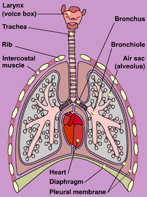
Oxygen is obtained from inhaled air. The air passes
down the trachea (windpipe) and enters the bronchi at the entrance to the lungs.
Each bronchus divides into smaller tubes called bronchioles. At the end of the
each bronchiole branch are sets of air sacs. These air sacs are called alveoli.
Oxygen from the inhaled air in the alveoli diffuses
into the blood. At the same time carbon dioxide from the blood plasma diffuses
back in the opposite direction. The alveoli have a very large surface area with
thin moist surfaces and an extensive supply of blood vessels. This allows rapid
gas exchange. Exhaled air passes back out through the trachea and air passages.
Only one third of the air contained in the lungs is exchanged during normal
breathing. The lungs only partially empty when you breathe out. Oxygen is
transported around the body in the blood by the circulatory system to respiring
cells where it enters by diffusion.
Inhalation
21% oxygen
1% other gases
78% nitrogen |
- Diaphragm contracts
- Ribs rise (intercostal muscles pull)
- Increased volume
- Air rushes in
|
Exhalation
17% oxygen
4% carbon dioxide and water vapour
1% other gases
78% nitrogen |
- Diaphragm relaxes
- Ribs fall (muscles relax)
- Decreased volume
- Air pushed out
- Exhaled air is warmer
|
| Alveolus |
The air sacs in
the lung are known as alveoli. Under the microscope they appear like a
bunch of grapes. |
| Bronchus |
The two bronchi
are passages that lead into the lungs from the central trachea. |
| Diaphragm |
A muscular sheet
found at the base of the lungs. Its contraction causes an increase in lung
volume on inspiration. |
| Intercostals |
The rib muscles
that cause raising of the rib-cage on inspiration. |
| Trachea |
Wind-pipe covered
in protective rings of cartilage that provide support. |
Respiration is the process used in all
living things for gaining energy. Energy is needed for keeping the vital organs
working and generally staying alive. It is also needed for moving muscles.
Keeping a constant body temperature is important to. Cell division, reproduction
and other important processes in the body all need energy to work.
Breathing in
>>> Inspiration
Diaphragm lowers and the ribs raise to increase the volume of the thorax. The
pressure decreases so air is drawn into the lungs.
Breathing out
>>> Expiration
Diaphragm raises and ribs lower which decreases the volume of the thorax. The
pressure increases and air is forced out of the lungs

Blood
is carried around the body in blood vessels called arteries and
veins. Exchange of substances occurs through tiny tubes called
capillaries.
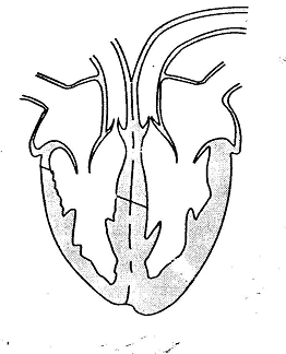
The
top chamber is called the atrium, and the bottom chamber is called the
ventricle. As you are looking at the heart diagram above, the right atrium is on
the left (imagine you are laying on the paper, it helps!). Deoxygenated (blue)
blood enters the right atrium from the body and is pumped to the lungs. The
oxygenated (red) blood enters the left atrium and is pumped around the body from
the left ventricle. Looking at it you can see how much more muscular this part
of the heart is, this is because the blood needs to be pumped all around the
body from here.
What you should be able to do.
- Investigate the effect of
exercise on your breathing rate.
- Use a chest model to show
how breathing takes place.
- Measure your chest size
during breathing.
- Test your exhaled air for
carbon dioxide with lime water.
- Compare the amount of
carbon dioxide in inhaled air with that of exhaled air.
- Test your exhaled air for
water with cobalt chloride paper.
- Know what artificial
respiration is how to apply it.
- Carry out an experiment
that shows the difference between fresh air and cigarette smoke.
- Design a leaflet for
primary school children to explain why they should not start smoking.
- List the main differences
between arteries, veins and capillaries.
- Plan an investigation to
see how your pulse and breathing are affected by exercise.
- Use a stethoscope to hear
the heart beating.
- List the things that can
increase the risk of heart attack and know how to reduce these risks.
- Observe and draw each
type of blood cell as seen under the microscope.
|